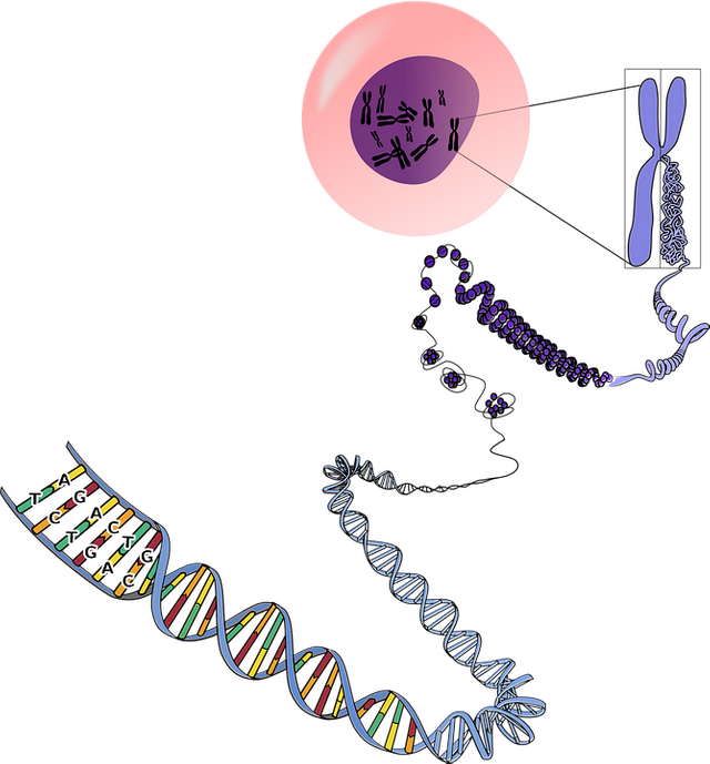
Source of image of pixabay
The interval that separates two successive divisions of a cell is named an interface and during this period it is said that the nucleus is interfásico, the duration of the interface is variable and depends on the textile to which the cell belongs. In the nucleus interfásico there can differ four constituent ones, the nuclear covering, the nucleoplasma, the cromatina and the nucléolo, in the eucariotas the nuclear content or nucleoplasma, it is separated from the citoplasma by a double membrane or nuclear covering.
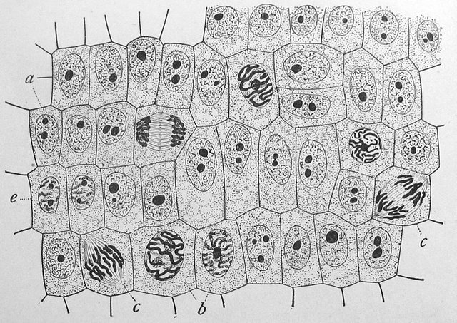
General sight of the Y-chromosomes his changeable aspect inside the cells: (a) Cell without splitting (there be observed the network of cromatina and the intensely dyed nucléolo); (b) nuclei prepared for the cellular division (it can be observed that the cromatina has become condensed); (c) Cells in different stadiums of division mitótica (it is possible to observe that the cromatina has stopped becoming condensed and the chromosomes have formed). In an apex of root of onion, observed with 800 increases, source of image of mastery of Wikimedia Commons, Author: Edmund Beecher Wilson.
The nuclear external membrane extends in the membrane of the reticulum endoplasmástico, in the nucleoplasma there can distinguish two called spheres nucléolos, which are usually next to a dense mass, which receives the name of cromatina. The nucléolos lack membranes and in them ARN is synthesized ribosómicos, also in the nuclei the newly synthesized ARNr they are packed by proteins and they lead to the ribosomas, the cromatina is formed by ADN associated with a group of structural proteins, the histonas that are the person in charge of packing the molecules of ADN inside the nucleus, there exist different increasing orders of empaquetamientos of the ADN, between which the simplest is the nucleosoma.
Every nucleosoma is formed by a protein nucleus constituted by several histonas, about which coils the ADN, the nucleosomas in turn are packed between them forming structure of major order. During the interface it takes place the processes of trascripción of the ADN and of synthesis of the proteins.
it is named a chromosome (of the Greek χρώμα, - τος chroma, color and σώμα, - τος soma, body or element) to each of the highly organized structures, formed by ADN and proteins, that contains most of the genetic information of an individual.
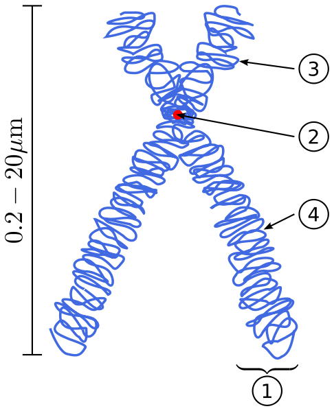
Schematic diagram of an already duplicated eucariótico and condensed chromosome (in metaphase mitótica). (1) Cromátida, each of the identical parts of a chromosome after the doubling of the ADN. (2) Centrómero, the place of the chromosome in which both cromátidas touch. (3) short Arm. (4) long Arm, source of image of mastery of Wikimedia Commons, Author:derivative work: Tryphon (talk) *Chromosome-upright.png: Original version: Magnus Manske, this version with upright chromosome: User:Dietzel65
In the cellular divisions (mitosis and meiosis) the chromosome presents his most well-known form, bodies well delineated in the shape of the Xth, due to the high grade of compactación and doubling, in the interface they cannot be visualized by means of the optical microscope of a clear way since they occupy chromosomal discreet territories. In the cells eucariotas and in you arch (to difference that in the bacteria), the ADN will always be in the shape of cromatina, that is to say associated strongly with a few named proteins histonas and not - histonas, the cromatina organized in chromosomes, is in the nucleus of the cells eucariotas and is visualized as a jumble of thin fibers, when it begins the process of doubling and division of the genetic material so-called (cariocinesis), this jumble of fibers initiates a progressive phenomenon of condensation that each of the chromosomes allows to visualize.
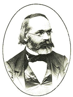
[Karl Wilhelm von Nägeli (Kilchberg March 27, 1817 - May 11, 1891) was a Swiss investigator of botany who discovered the chromosomes in the XIXth century, source of image of mastery of Wikimedia Commons, Author:The original uploader was Sixat at German Wikipedia.]
The nucleus in division
Contains all the information necessary for the functioning of the cell, the cellular division is the phenomenon for which a cell mother of origin to two cells daughters with the same genetic information. Before of begins the cellular division the ADN, it doubles and the cromatina is hydrated and becomes condensed leading to the formation of the chromosomes mitóticos.
The cromatina of nuclei in interface, when it is observed by means of skills of electronic microscopy, it is possible to describe as a necklace of accounts or a rosary, in which every account is a spherical or globular subunit that is named nucleosoma; the nucleosomas are joined between yes by means of fibers of ADN. One continues, then, that the basic unit of the structure of the cromatina is the nucleosoma. A typical nucleosoma is associated at 200 base par (pb) of ADN and is formed by a marrow (core in English) and a ligador (or linker), the marrow is formed by an octámero constituted by two subunits of the histonas H2A, H2B, H3 and H4.
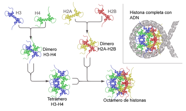
Structure of the nucleosoma, source of image of mastery of Wikimedia Commons, Author:Asasia
The number and the form of the chromosomes are typical of every species, new paragraph has form of bastoncitos with a contrition, call centrómero, which divides two arms, often on the arms of the chromosome appears a strangulation named constriction secondary, at the beginnig of the cellular division the nucléolos are joined to the constriction secondary of some chromosomes. Also it usually appears in one of the ends of the chromosomes, a circular formation called satellite, which is joined to the chromosome by a filament, the majority of the cellular nuclei contain pairs of chromosomes, so called homologous chromosomes to the chromosomes (n), it is typical of the species and every nuclei it contains two I play identical of n chromosomes.
Bibliography
Essentials of basic biology. - Page 86 for Cherry tree García, Miguel - 2013,
evolutionary Biology - Page 88 for Raúl Montenegro - 2001.
THOMPSON * THOMPSON. Genetics in medicine Student Consult for Robert L. Nussbaum, Roderick R. McInnes, Huntington F Willard - 2008.
