Throughout the covid pandemic and its aftermath the American doctor Tom Cowan has been one of the most outspoken critics of the science and practice of modern virology. In a recent video presentation with two other virus skeptics, Sam and Mark Bailey, Cowan contrasts the failure of virologists to isolate viruses with the success of chemical analysts to isolate minuscule traces of proteins and other substances from given samples:
... the question of whether one could purify something, meaning have only something the size and complexity of a virus ... because there could be things in the world that exist that we simply don’t have the technology—the techniques—to actually isolate and purify. Now, that’s why some of us have brought up things like Stefan’s giant viruses, which are actually spores, or bacteriophages, which are actually break-down products of bacterial cultures, because, and it should be very clear to everybody: this is not a technical problem. The ability to purify something the size and complexity of a virus is not a technical problem. I’ve run this by analytical chemists, and I’ve said: “Here’s the size, here’s the complexity.” They say they could isolate and purify it in a matter of days. Every single virus on the planet. So it’s not a technical problem. And when I said, “Well, why do you think they haven’t been able to isolate, ie purify, this virus so they could study it?” they said, “It’s simple: it’s not there.” Because that’s the only other reason, if it’s not a technical problem. It’s not because there’s too little of it. There are ten million in a sneeze. I asked this Australian analytical chemist: “If you put ten million particles in a sneeze the size of a virus, the complexity of a virus, could you find it?” And he said, “Of course!” So, this business of “Well, we can’t find it, there’s not enough, blah, blah, blah,” ... at the end of the day there’s only one reason: It’s not there. And if it’s not there, then all the RNA or DNA sequences ... you have no idea where they’re coming from. (Timestamp: 33:45-35:52)

Mark Bailey then corrects Tom:
Just one slight correction there, Tom. Apparently it’s 200 million SARS-CoV-2 particles per sneeze. (Timestamp: 35:53-36:02)
Mark then discusses the isolation of molecular substances like insulin and other protein or protein-like substances. These are orders of magnitudes smaller than viruses but are routinely isolated:
Now, insulin is a protein. Now, when we were treating diabetics, you know, in acute situations, when, say, their blood sugar was four or five times the upper limit of normal, we wouldn’t accept someone giving us a sample of insulin saying, “Well, sorry, I don’t really know what the purity is here.” I mean, the samples we were using of insulin—I can’t remember from memory—but I know that they’re in the mid- to high nineties in terms of their purity. And they have very predictable effects. Like, when they give you a unit of a particular type of insulin, it’s got predictable effects on the blood sugar level. So, again, like you say, Tom, here’s an example of a particle that is purified, and that’s a tiny, you know, in the scheme of things, that’s just a protein, just one protein by itself. (Timestamp 36:25-37:22)
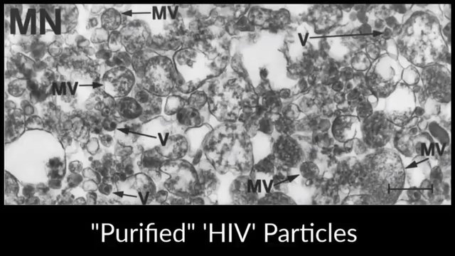
Mark then displays an electron micrograph which, it is claimed, shows the purified and isolated HIV virus. MN = purified HIV-1(MN)/H9, containing some mature viruses (V) and numerous nonviral particles—presumably microvesicles (MV). The measuring bar represents 1 μm:

It looks like a Picasso painting. (Timestamp 37:48-38:06)
If pathogenic viruses such as SARS-CoV-2 exist and produce diseases in humans and other animals, then surely it should be possible to isolate them from infected bodily fluids in the same way that chemical analysts isolate traces of specific chemicals. Viruses are tiny compared to bacteria, but they are huge compared to molecules. Moreover, they are of a similar size and structure to bacteriophages, which have been successfully isolated and purified by following a simple and obvious procedure. It ought to be child’s play to isolate pathogenic viruses from sick people by following a similar procedure:
Take samples of mucous, lung fluid, pus, etc from a sick person.
Place them in a centrifuge to separate out the constituents according to their masses.
Pass the lighter component—the supernatant—through fine filters to remove the larger particles.
Examine the resulting isolates under an electron microscope and see if you have truly isolated the viral particles.
Infect someone or a test animal with the isolates to see whether they become ill.

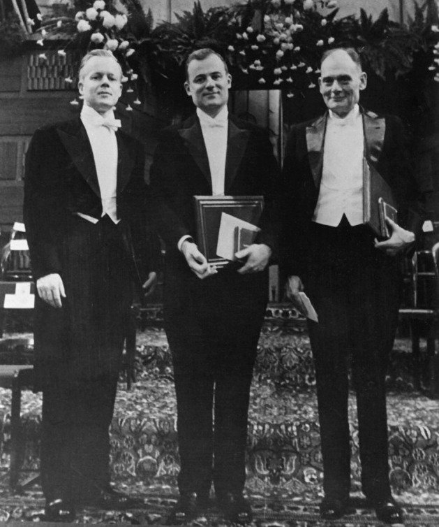
In a podcast he made in February 2022 with health consultant Ben Greenfield, Tom Cowan alleged that virologists attempted to do just this in the 1930s and ’40s, but they failed to isolate any viruses and no test animals became sick. So, instead, they developed a complicated method to “isolate” viruses by growing them in specialized tissue cultures and observing the so-called cytopathic effect. In 1949 John Franklin Enders, Frederick Chapman Robbins and Thomas Huckle Weller succeeded in culturing the alleged polio virus in a laboratory setting. In 1954 the three shared the Nobel Prize in Physiology or Medicine “for their discovery of the ability of poliomyelitis viruses to grow in cultures of various types of tissue” (The Nobel Prize). This became the standard method of “isolating” pathogenic viruses and has remained so to this day.
Ben: And so, what a virologist would do if they did want to like isolate and characterize and demonstrate the existence of a virus or a new thing like SARS-CoV-2, they would take a bunch of samples from infected people like blood, or sputum, or I don’t know what else you might collect. Some type of bodily secretion from people who are demonstrating symptoms of what we suspect might be a virus, and those symptoms are unique and specific enough to where we can say, okay, all these people are showing signs of something that shows that they have something that we suspect might be something. And then, basically, what they then do is they take those samples, and ideally, they would not mix them with anything else that has any other genetic material, and then they would do what they do in the lab, filter it and macerate it and centrifuge it and purify this specimen. And, that would be a common virology technique that I understand the virologists have done for quite some time to isolate bacteriophages and so-called viruses. And, if they did that, that would allow them to demonstrate—with then typically an electron microscopy tool—a whole bunch of different particles that they would then be able to say are the isolated and purified virus. Then, they could go and check those with microscopic techniques to determine the purity and to further characterize the particle examining the structure, or the chemical composition, or what we call the morphology. And then, you’d have to extract the genetic material from that from these purified particles. And then, that would be done using a genetic sequencing technique. That’s old technology, it’s been around for decades. And then, you would analyze whether those particles are outside the cell, what we call exogenous. Meaning that they were something that originated from somewhere outside the human body. And, they weren’t the normal breakdown products of dead or dying tissue in the body. (Tom Cowan Podcast: 19 February 2022)

And, if you’ve done all of that, you would have fully isolated and characterize and genetically sequence this particle that you would then want to call a virus. And then, if you successfully did that, you’d then have to go out and show that it’s actually causally related to a disease, so you’d have to expose a bunch of healthy subjects. Preferably these days, they would be cute little animals and you’d expose them to this isolated purified thing that you suspect is a virus in the manner in which you thought that virus might be transmitted, whatever, saliva, or cough, or I don’t know, blood transfusion, or whatever. And then, if those animals got sick with the same disease as the original ones you were studying, if you were to confirm that with some type of clinical finding or autopsy finding, then you would have the bodies in the streets and you would say, okay, we’ve isolated something we’re going to call the virus that actually causes the disease. And we’ve demonstrated infectivity and transmission in addition to the existence of such a virus. That’s what you would ideally do, right?
Thomas: That’s not ideally. That’s the only way to do this in any rational way. Ben, you got it exactly right. The only thing I would add is all of these things you’re talking about as you said are standard virology techniques that go back to the ’30s.
Ben: Okay, okay.
Thomas: If somebody says to you, “Oh, well, you can’t find this purified virus”–in 1940, there’s pictures of bacteriophages which are identical shape and morphology as viruses essentially and they easily found that. And, the only other thing I would say is when you do that, you should find the something that’s millions of copies of identical particles, not like one is this size and one is that size. (Tom Cowan Podcast: 19 February 2022)
Tom’s final point is one that is often overlooked. In electron micrographs that purport to show isolated viruses, the viruses are always of different sizes—and sometimes even of different shapes. Even on Wikipedia SARS-CoV-2 is described as being 60–140 nanometres ... in diameter. But if a virion infects a cell, takes over the machinery of that cell, and replicates itself using its own genome as a template, then all the resulting virions should be clones of the original: as identical to it as identical twins are to each other. And viruses are not alive, so they cannot grow or alter their morphology after they have been created—something identical twins can do—so they should remain absolutely identical to one another for the whole of their existence.
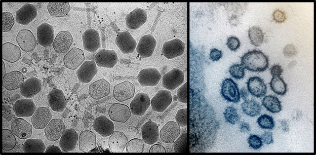
Compare the following two images: the one on the left is a transmission electron micrograph (TEM) of bacteriophages, while the one on the right is alleged to be a TEM of SARS-CoV-2. The bacteriophages do indeed look identical—but the viruses?

And that’s a good place to stop.
References
- Julian W Bess, Jr, et al, Microvesicles Are a Source of Contaminating Cellular Proteins Found in Purified HIV-1 Preparations, Virology, Volume 230, Issue 1, Pages 134-144, Academic Press, Cambridge, Massachusetts (1997)
- John Franklin, Thomas Huckle Weller, Frederick Chapman Robbins, Cultivation of the Lansing Strain of Poliomyelitis Virus in Cultures of Various Human Embryonic Tissues, Science, New Series, Volume 109, Number 2822, Pages 85–87, American Association for the Advancement of Science, Washington, DC (1949)
Image Credits
- COVID-19 Poster: © 2021 Dublin Region Homeless Executive, Fair Use
- Tom Cowan: © Tom Cowan, Fair Use
- Purified HIV Particles: Julian W Bess, Jr, et al (transmission electron microscopists), © 1997 Academic Press, Fair Use
- Thomas Weller, Frederick Robbins & John Enders: Francis A Countway Library of Medicine, Harvard Medical School, Fair Use
- Ben Greenfield: © Ben Greenfield Life, Fair Use
- Bacteriophages: © ZEISS Microscopy, ZEISS Libra 120 TEM, Creative Commons License
- SARS-CoV-2: National Institute of Allergy and Infectious Diseases, Rocky Mountain Laboratories, Hamilton, Montana, © NIAID-RML, Creative Commons License
Online Resources
- Ben Greenfield Podcast, 19 February 2022
- Compilation Video of Tom Cowan on Viruses
- ViroLIEgy
Stefan Lanka
