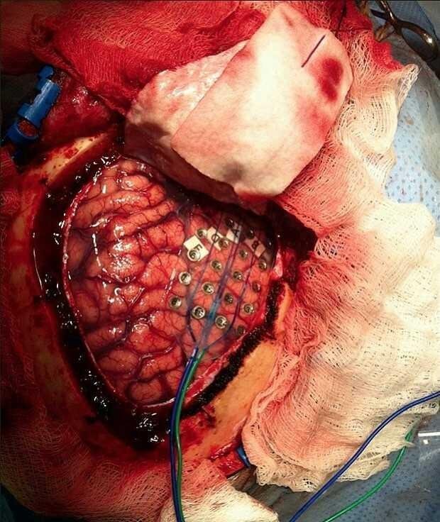
This image demonstrates how Electrocorticography (ECog) monitoring is being done.
A 32-contact grid is placed over the right temporal lobe, just over the areas with interictal epileptiform discharges.
Electrocorticography (ECoG) is performed by using electrodes placed directly on exposed cortical surface to record brain electrical activity. This is considered an invasive procedure, simply due to the fact that craniotomy is needed in order to expose the brain and directly place the electrodes, done either intraoperatively or extraoperatively.
ECoG has been used to localize epileptogenic zones during presurgical planning, map out cortical functions, and to predict the success of epileptic surgical resectioning.
An ECog recording done in a neurosurgical patient who suffers from epilepsy raises the exact problematic area that contain abnormal neuronal pattern that is responsible for the seizures. This active seizure focus can then be surgically resected without further complications.