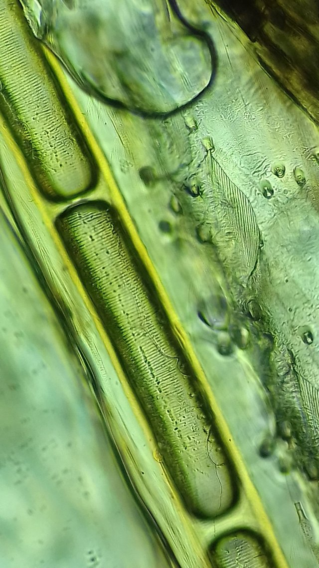
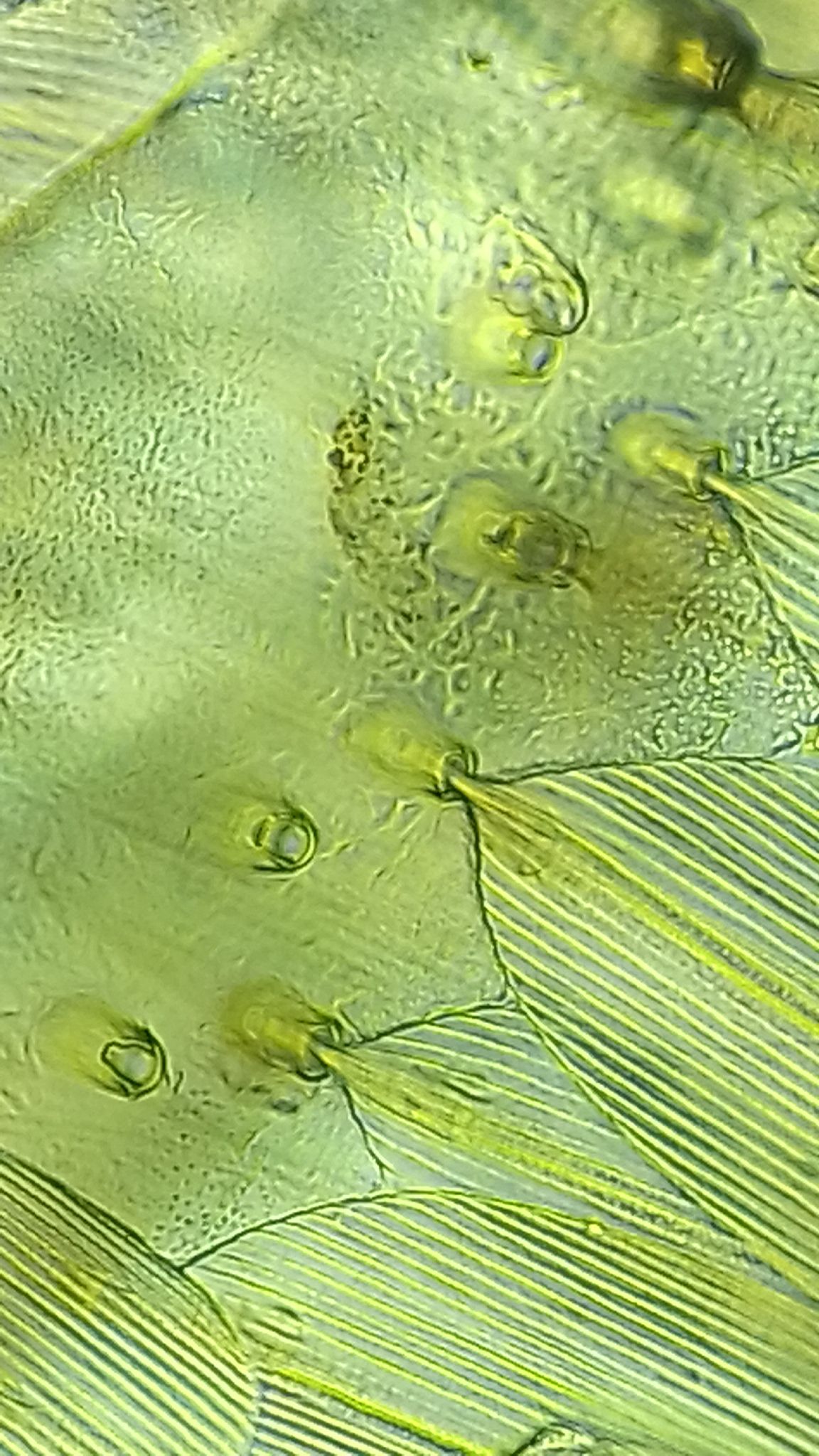
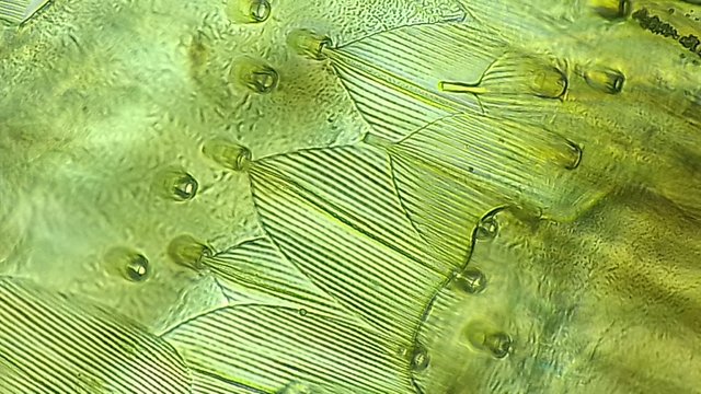
Beginning from the top downwards,
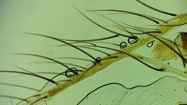
The first three picture are of the contents of a vein within a Mayfly wing and the very tiny feathers that cover the surface of a common moths wings, each feather fits into an individual socket and can more around as the insect is in flight, these act as dampeners to prevent them being detected and keep the insect insulated in the colder evenings.
The next two pictures are of pollen grains found in our local honey, magnified 100 times.
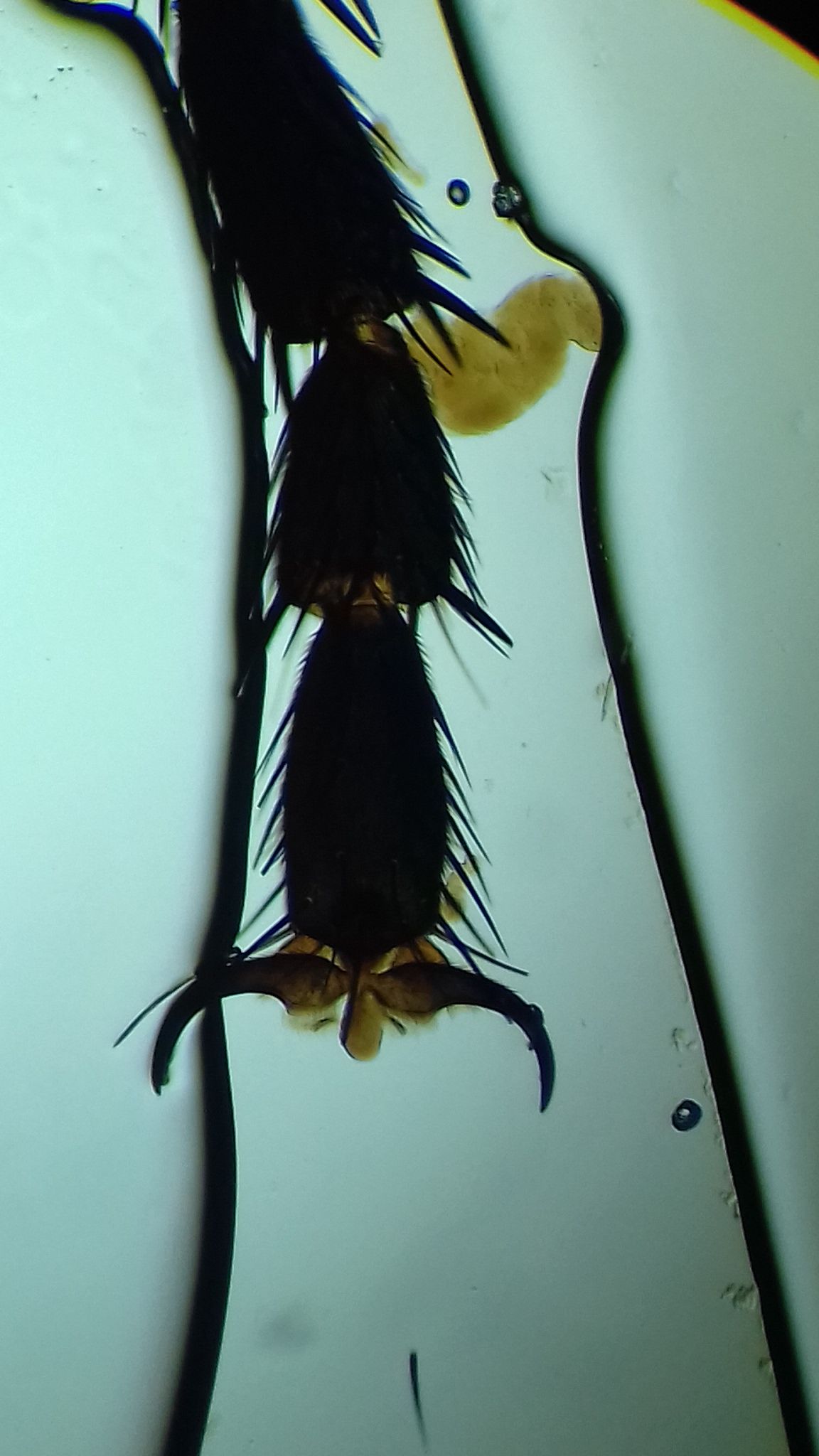
Next is the claw found on the end of a bluebottle leg, they also have added tines or spurs to enable them to grip the smallest anchor point.
Next are the minute hairs found upon the surface of the Mayfly wing and the lymph inside the tiny veins.
Next the same Mayfly wing and a single hair in its socket showing the folicle.
Bottom is an individual moth feather taken in dark field mode.
![20180315_221233[1].jpg](https://steemitimages.com/DQmeeASDUMr8emPiR3TZq5rbSHGtsySMq62zd5U3kCpJAiZ/20180315_221233%5B1%5D.jpg)
![20180315_222500[1].jpg](https://steemitimages.com/640x0/https://steemitimages.com/DQmZoKrksnrS6VNXHVsAaeCdBdoX1AtwA7hcdYh9fRETVKX/20180315_222500%5B1%5D.jpg)

Below is a section from a fly compound eye, notice each individual Hexagonal lenses in the matrix.
![20150101_000550[1].jpg](https://steemitimages.com/640x0/https://steemitimages.com/DQmZ7r8tfY5VdnVx9MreFYBv6GdojHhNbQv7Yv3iigfGV5x/20150101_000550%5B1%5D.jpg)
Below showing the Exoskeleton and leg joint of a house fly, with its protective spines at the joint area.
Seen below is what a tiny drop of saliva looks like after a meal, notice the long starch fibres and the millions of other particles in food as it is digested by carbohydrates in the saliva.
Below is the follicle of a human hair and shaft.
The pictures were taken by holding my mobile phone up to the microscope eyepiece, if your steady enough you can get decent enough photo's, begin by centralizing the camera in the lens then slowly move inwards until the camera focuses, once you get the hang of it it becomes much easier, take several pictures at once just incase things move.
Thanks for looking.