Hi welcome to my 1st blog about biological science! It's a random feeling of wanting to share this laboratory experiment we had in our BIO 108: Molecular Cell Biology, it's the easiest and simplest way to see and appreciate our cells under the microscope. It was just so amazing and interesting how these little structures are existing and actually part of our being.
Staining is a technique used to enhance visualization of structures in biological tissues under a microscope. Stains then are salts that color particular ions in the bacterial cell and make more visible distinction. The chemical composition of the cell determines which stain is absorbed. Acidic parts of a cell absorb stains that are positively charged. Staining can also be carried out to highlight metabolic processes or to differentiate between live and dead cells in a sample.
Before tissues are stained, a thin layer of cells that have been sliced from specimen (a smear ) is prepared by fixing. Fixing a specimen that has been placed on a slide is done by either allowing it to any at room temperature, or by passing the specimen quickly over a flame. Next the specimen is stained either one of the two categories: 1) Morphological stains to provide information about the cell size, shape, and arrangement. Examples of morphological stains include the simple stains and the negative stain. 2) Differential stains to differentiate bacteria based on the chemical composition of the cell wall. The differential stains require two stains (primary stain and counterstain) be used; one for the positive bacteria, the other for the negative bacteria. The differential stain that will be used in this activity is Gram stain.
Many staining techniques require you to start by carrying out a smear preparation first. A smear is done to fix a thin layer of cells to the microscope slide prior to staining cells must adhere to the slide so they not wash off during the staining and washing processes. There must only be a thin layer of cells so the morphology and arrangement of individual cells can be visualize.
Materials:
A. Eyepiece micrometer; Chemicals: Methylene blue stain
B. Chemicals: Crystal violet stain, Grams iodine, 95% ethanol, Safranin stain
Lets get it started!
Procedures:
A. Simple Staining
- Add a few drops of methylene blue to cover the whole smear prepared earlier. Let it stay for one (1) minute.
- Wash off the stain by rinsing it with water. Do not apply water directly above the slide, just let if flow into your smear. Blot dry with tissue at the slide.
- Examine under the microscope from the lower power up to the high power objectives.
- Record your observations through drawings or photo documentation.
Photos:
Scanner objective
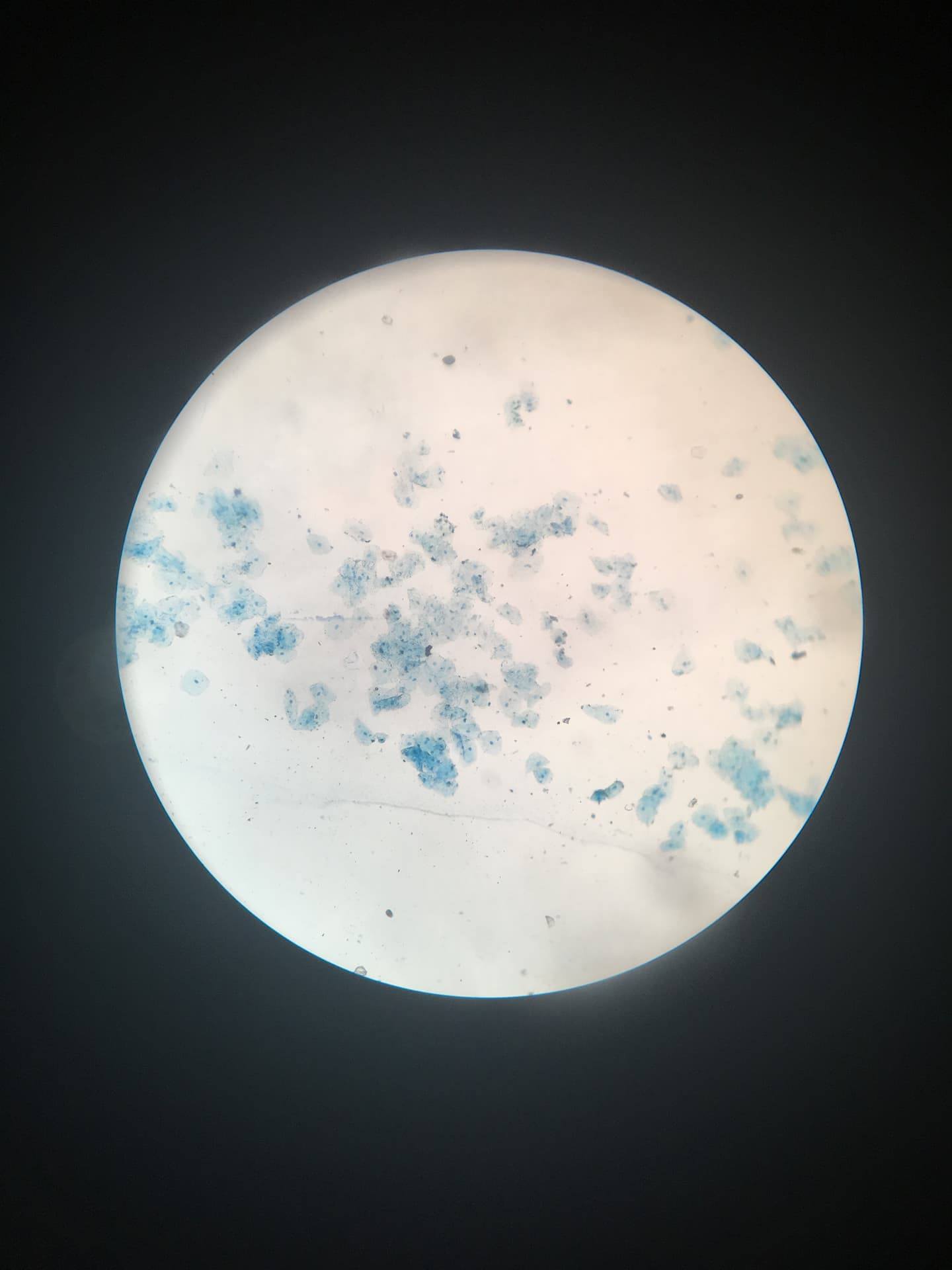
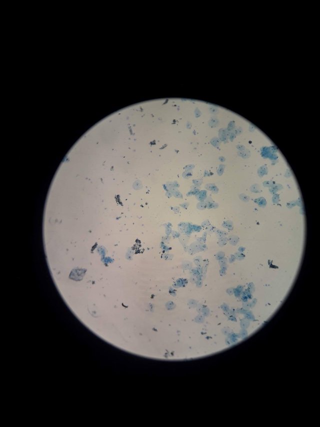
Low power objective
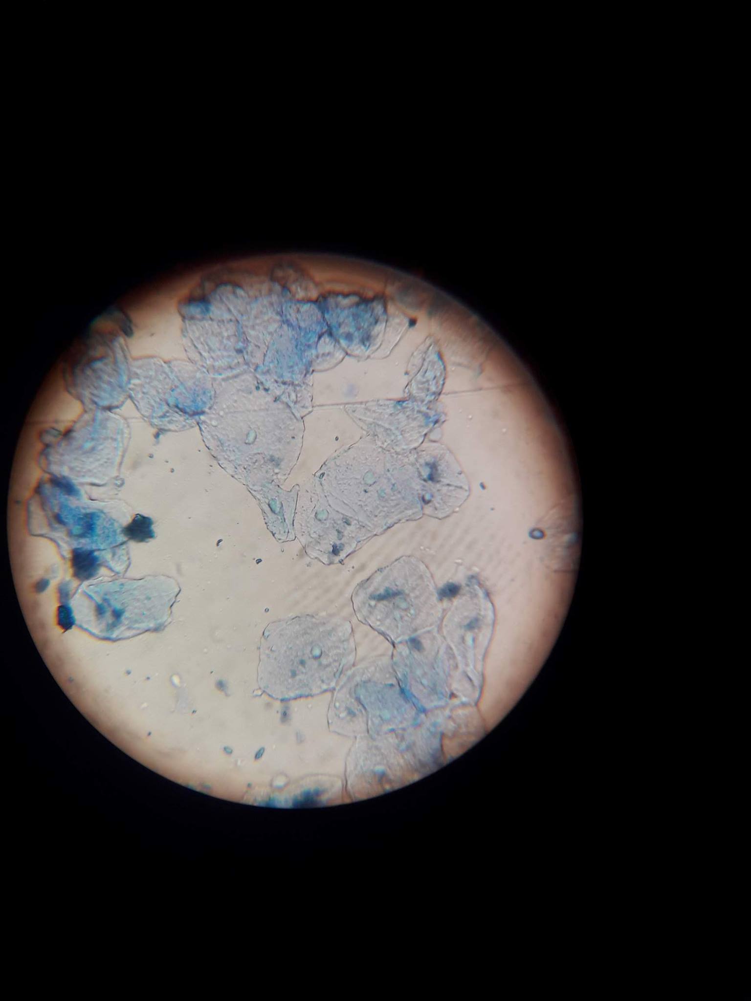
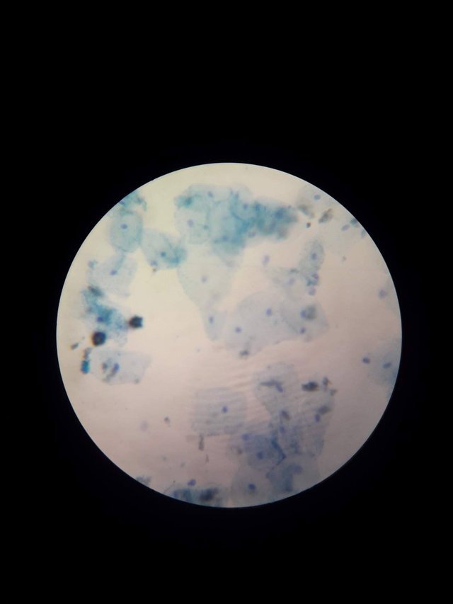
As you can see, with the help of the methylene blue stain you can see immediately the nucleus of the cell which is the darkened part in the middle of every irregular shapes. These are actually your cheek cells! Imagine how your cheek inside your mouth contains a millions of this. Amazing!
B. Special Staining: Gram Staining
- Prepare smear, but this time obtain sample materials lodged between you teeth or on the surface of your teeth at the gum line.
- Add a few drops of Gram I (crystal violet) into your smear. Do not drop it directly into your smear. Let it flow towards the slide.
- Let the stain stay for 1 minute. Then wash all the stain off with water.
- Add a few drops of Gram II (Gram's iodine) by letting it flow into your smear and then wait for 1 minute. Wash off with water.
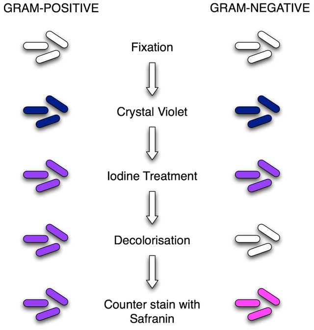
- Decolorize your smear with Gram III (ethyl alcohol) by letting it flow briefly for 30 seconds into your slide. Quickly wash with water thereafter.
- Lastly, stain the smear with Gram IV(Safranin) for 1 minute.
- Wash all stain off with water and blot dry.
- Observe under the microscope. Determine the shape, arrangement of the cells, and the Gram reaction. Record your observations through photo documentation.
Photos:
The solutions after washing off the stains in the smear
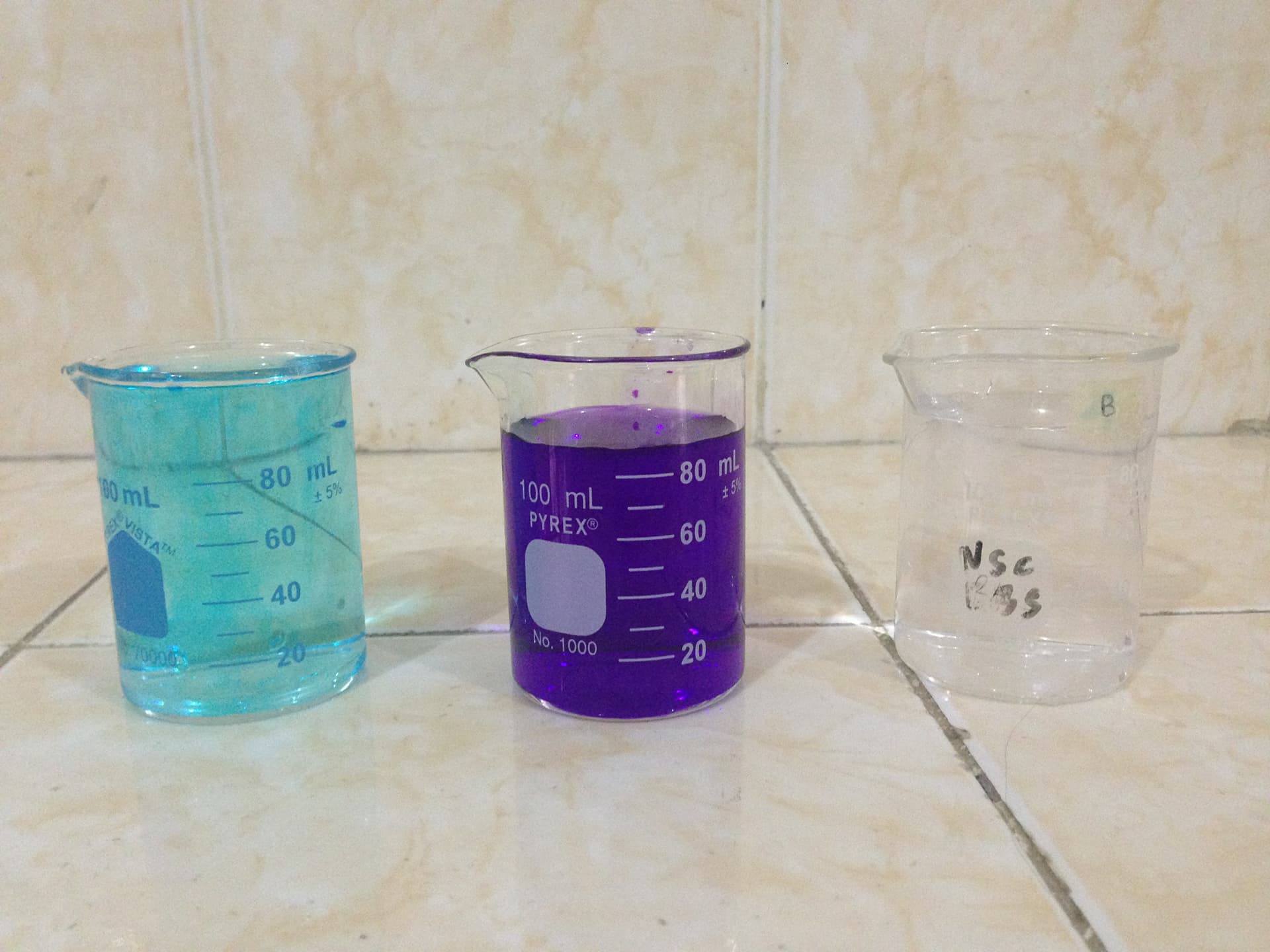
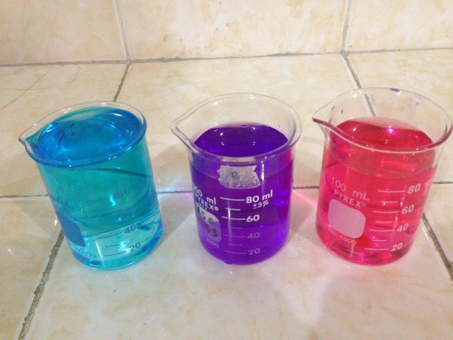
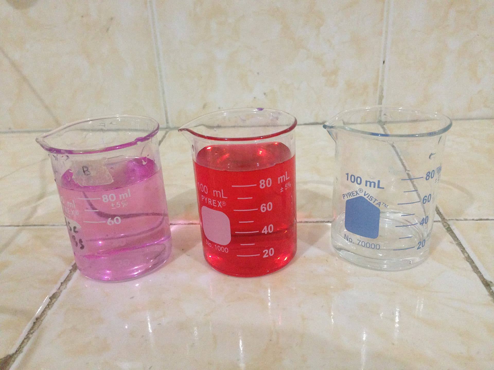
For an easy way to distinguish the bacteria to their color (since I haven't documented them during our lab hour) here's a photo : [Source] (https://microbeonline.com/gram-staining-principle-procedure-results/)
And here's our final result! Our professor was so glad since only me and my groupmates were able to see the bacteria under the microscope.
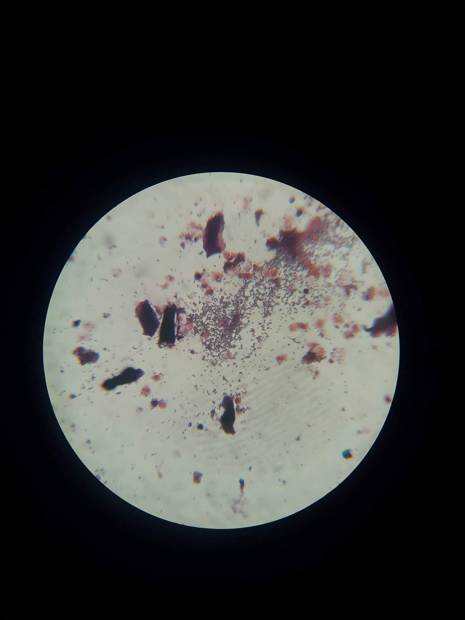
With just a simple lodging of your teeth between your gum line, imagine this number of bacteria (specified by the encircled part of the photo) present and multiply it by nth times! Oh no!

And now let me ask you again, are you sure your mouth is healthy? Hahahahaha!
I would like to thank @welcoming for suggesting me to watch posts of @sco I feel inspired in making science posts then on. Thank you @theaustrianguy
References:
Bruckner, M. Z., Microbial life educational researchers: Basic Cellular Staining
Chan, E.C.S., Pelczar, M. Jr. and Kreig, N.R. 1993. Laboratory Exercises in Microbiology 6th ed.
Devlin, K.R. Microbiology Laboratory Exercises
Kaila ko ni! Proud of you jane! 😘
Downvoting a post can decrease pending rewards and make it less visible. Common reasons:
Submit
Hahahaha avaad! Thanks ya Prinss!:*
Downvoting a post can decrease pending rewards and make it less visible. Common reasons:
Submit
This deserves to be curated. Very informative. Our group is proud to have you. #steemitachievers
Downvoting a post can decrease pending rewards and make it less visible. Common reasons:
Submit
Thank you for appreciating my post @lebron2016!
Downvoting a post can decrease pending rewards and make it less visible. Common reasons:
Submit
feel nako ma curie ka amegs hahahaha go janeyyyyy bet sa oks
Downvoting a post can decrease pending rewards and make it less visible. Common reasons:
Submit
Friend nako ni <3 hahahaha gogogo janey
Downvoting a post can decrease pending rewards and make it less visible. Common reasons:
Submit
feel nako busy pud ka run? hahaha
Downvoting a post can decrease pending rewards and make it less visible. Common reasons:
Submit
hahahaha naa lang koy apilan nga contest po :D HAHAHAHA
Downvoting a post can decrease pending rewards and make it less visible. Common reasons:
Submit
hahahaha lrj ang competition karon hahaha
Downvoting a post can decrease pending rewards and make it less visible. Common reasons:
Submit
Hahahah cheering kyka ha pro wa koy upvote: ( Hahahaha mwaa
Downvoting a post can decrease pending rewards and make it less visible. Common reasons:
Submit
Congratulations @janeynarzoles1! You have completed some achievement on Steemit and have been rewarded with new badge(s) :
Click on any badge to view your own Board of Honor on SteemitBoard.
For more information about SteemitBoard, click here
If you no longer want to receive notifications, reply to this comment with the word
STOPDownvoting a post can decrease pending rewards and make it less visible. Common reasons:
Submit