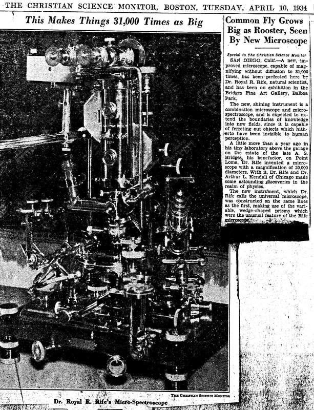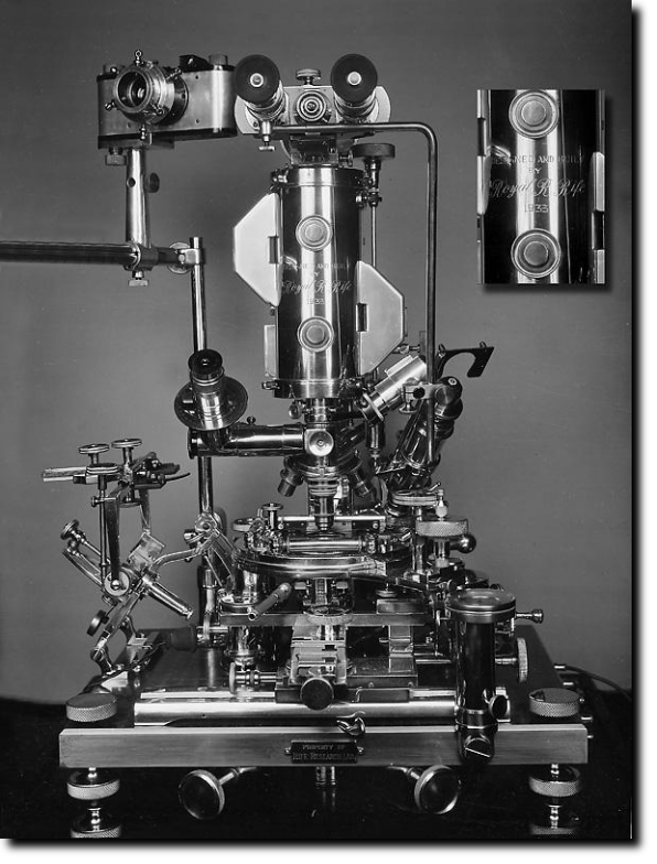
"Royal Raymond Rife was the inventor of the Universal Microscope which he presented to the world in 1933. Besides being the most powerful optical microscope ever made up to that time, it was also the most versatile. The Universal used all types of illumination: polarised, monochromatic or white light, dark field, slit ultra and infra-red. It could be used for all manner of microscopical work, including petrological work or for crystallography and photomicrography. According to a report submitted to the Journal of the Franklin Institute it had a magnification of 60,000x, and a resolution of 31,000x. The ocular of this instrument was binocular, but it also had a detachable segment lower in the body for monocular observation at 1800x (x=power) magnification.
One of the most attractive features of this microscope is that, in contrast to the Electron Microscope, the Universal Microscope does not kill the specimens under observation and affords observation of natural living specimens in all circumstances, meaning it does not rely on fixing or staining to render visibility or definition.
Rife achieved this by using various modes of lighting to bring virus into visibility in their natural colors."
https://www.rife.de/brief_history.html

"What Royal Rife built initially was his "Universal Prismatic Microscope". This incredible microscope amplified his view of LIVE SPECIMENS by up to 60,000 times! This allowed him to view live pathogens in controlled environments. Rife was the FIRST PERSON in the world to actually see (and photograph) live viruses! Rife found a means to building this microscope with a very long optical path with specially-ground quartz prisms in the barrel of his microscope. In addition, he determined that white light, which contains all wavelengths of visible light, was not suitable for high-magnification optics, and used a "Risley" prism between his light source and the sample, thus illuminating the sample with a single frequency of monochromatic light (very similar to high-definition research microscopes of today which use lasers).
Royal Rife's microscope was a stunning advance. Unlike the electron microscope, Rife's microscope made it possible to study living bacteria, viruses, fungi, and so forth. An electron microscope kills it's specimens. Royal Rife's remarkable breakthrough used a new approach to bend light. As an end result, Rife was able to prove that bacteria could change their form (or die), because he and others could literally watch it happen!"
https://www.royal-rife-machine.com/
"The light, which contains the carrier and a mixture of selected signals in the UV range, stimulates the biological material in the Somatoscope to the point that the specimens give off their own light. (Rife referred to this as luminescence.) This is the key to the ultra-high resolution that has been achieved by Gaston Naessens."
"The Somatoscope does not attempt to illuminate the specimen by passing light through two small objects. Instead, the illumination source is actually stimulating the specimen to the point it generates its own light. The light itself expands as it moves outward and because the specimen itself is generating the light, the physical restrictions encountered by regular optical microscopes do not apply. By converting the specimen into a light source, Gaston Naessens has converted the magnification problem from one of resolution to that of light detection! At magnification levels above 5,000 diameters, light levels drop off the point that film is necessary, but the resolution is there."
http://www.rexresearch.com/naessens/naessens.htm
Royal Rife Microscope shatters Cancer.m4v
"The story of Royal Rife and his place in history are still up for discussion. This is the very rarely seen footage of his microscope and the shattering of cancer cells with frequencies."