SPACE FORCE HEALTH UPDATES
DAY 1
SENSES OF THE BODY
LOGO

MOTO: On the wings of superstars we are health and safety
I am glad to be the first official writer in the health department after and introductory post by my leader @peterakpan see link below
https://steemit.com/steemjet/@peterakpan/update-from-space-force-health-department
Without the special senses global steem adoption can't be ascertained
HEARING AND THE EAR
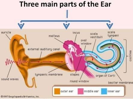
The ear is the organ of hearing. It is supplied by the eight cranial nerve, i.e., the cochlear part of the best I bulk cochlea nerve which is stimulated by vibrations caused by sound waves. With the exception of the auricle (pinna), the structures that form the ear are encased within the petrous portion of the temporal bone.
STRUCTURE OF THE EAR
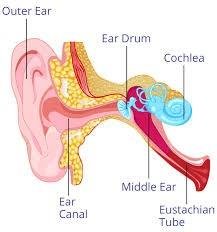
The ear is divided into three distinct parts
External ear
Middle ear (tympanic cavity)
Internal ear
🌘 EXTERNAL EAR
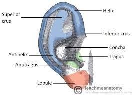
The external ear consist of the auricle (pinna) and the external meatus.
THE AURICLE
The auricle is the expanded portion projecting from the side of the head. It is conposed of fibroelastic cartilage covered with skin. It is deeply grooved and ridged and the most prominent outer ridge is the helix.
EXTERNAL ACOUSTIC MEATUS
This is slightly "S" - shaped tube about 2-5 cm long extended from the auricle to the tympanic membrane (ear drum). The lateral third is cartilagibous and the remainder is a canal in the temporal bone. The meatus is lined with a thin layer of skin, continous with that of the auricle. There are numerous celeminous glands in the skin of the lateral third. This are modified sweat glands that secret cerumen (wax), a sticky material containing lysosome and immunoglobulins. Foreign materials, e.g., dust, insects, and microbes, are prevented from reaching the tympanic membrane by wax towards the exterior.
🌘 TYMPANIC CAVITY OR MIDDLE EAR
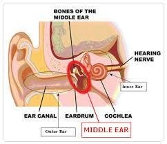
This is an irregular-shaped cavity within the petrous portion of the temporal bone. The cavity, its contents and the air sacs which opens oit of it are lined with mucous membranes. Air fills the cavity, reaching it theough the pharyngotympanic (auditory) tube which extends from the nasopharynx. It is about 4cm long and is lined with ciliated epithelium. The presence of air at atmospheric pressure on both sides of thr tympanic membrane enables it to vibrate when sound waves strike it.
Auditory ossicles
These are three very small bones that extends across the cavity from the tympanic membrane to the oval window. They form a series of movable joints with each other and with the medial wall of the cavity at the oavl window. They are; the malleus, incus, and stapes
🎳 The malleus
The malleus is the lateral hammer-shaped bone. The handle is in contact with the tympanic membrane and the head forms a movable joint with the incus.
🎳The incus
The incus is the middle anvil-shaped bone. Its body articulates with the malleus, the long process with the stapes, and it is stabilised by the short process, fixed by fibrous tissue to the posterior wall of the cavity.
🎳The stapes
The stapes is the medial stirrup-shaped bone. Its head articulates with the incus and its base fits into the oval window.
The three ossicle are held in position by fine ligaments.
🌘 INTERNAL EAR
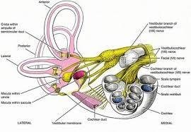
The internal ear contains the organ of hearing and balance and is generally described in two part, the bony labyrinth and the membranous labyrinth.
🔥Bony labyrinth
This is a cavity within the temporal bone lined with periosteum. It is larger than the membrane labyrinth of the same shape which fits into it, like a tube within a tube. The space between the bony walls and the membrabous tube is occupied by perilymph. The membranous labyrinth also vontains fluid, the endolymph.
The bony labyrinth consist of:
vestibule
cochlea
semicircular canals
🔥Membranous labyrinth
The membranous labyrinth is the same shaped as its bony counterpart and is separated from it by perilymph. It contains endolymph. It is divided into the same parts: the vestibule which contains the uricle and saccule, the cochlea and three semicircular canals.
PHYSIOLOGY OF HEARING
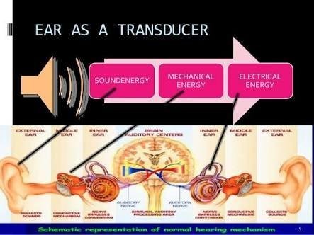
Every sound produces sound waves or disturbance in the air, which travel at about 332 meters (1088) feet per second. The auricle, because of its shape, concentrate the waves and directs them along the auditory me at us causing the tympanic to vibrate. Tympanic membrane vibrations are transmitted through the middle ear by movement of the ossicles. At their medial end the footplate of the stapes rocks to and fro in the oval window, setting up fluid waves in the perilymph. These indent the membranous labyrinth and the wave motion in he endolymph stimulates the neuroepithelial cells of the organ of corti. The nerves impulse produces pass to the brain in the cochlear portion of the eighth cranial nerve (VII). The fluid wave is finally expended into the middle ear by vibration of the membrane of the round window. This nerve, the best I bulk cochlea nerve, transmit the impulses to various nuclei in the pins varolli and midbrain. Some of the nerve fibers pass to the hearing area in the cerebral cortex where sound is perceived.
SIGHT AND THE EYE
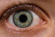
The eye is the organ of the sense of sight situated in the orbital cavity and it is supplied by the optic nerve (second cranial nerve). It is almost spherical in shape and is about 2.5 cm in diameter. The space between the eye and the orbital cavity is occupied by fatty tissue. The bony walls of the orbit and the fat help to protect the eye from injury. Structurally the two eyes are separate but, unlike the ear, some of their activities are vo-ordinated so that they function as a pair. It is possible to see with only one eye but three-dimemtional vision is impaired when only one eye is used, especially in relation to the judgement of distance.
STRUCTURE OF THR EYE
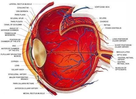
There are three layers of tissue in the walls of the eye.
They are:
The outer fibrous layer: sclera and cornea
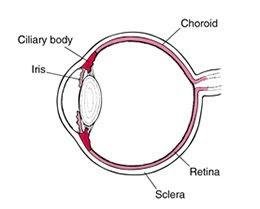
The middle vascular layer: choriod, ciliary body, and Iris
The inner nervous tissue: retina
🌘 SCLERA AND CORNEA
The sclera, or white of the eye, forms the outermost layer of tissue of the posterior and lateral aspects of the eyeball and is continuous anteriory with the transparent cornea. It consists of a firm fibrous membrane that maintains the shape of the eye and gives attachments to the extraocular or extrinsic muscles of the eye. Anteriory the sclera continues as a clear transparent epithelial membranes, the cornea. Light rays pass through the cornea to reach the retina. The cornea is convex anteriory and is involved in refracting or bending light rays to focus them on the retina.
🌘 CHOROID
The choroid lines the posterior five-sixeths of the inner surface of the sclera. It is very rich in blood vessels and is a deep chocolate brown in colour. Light enters the eye through the pupil, stimulates the nerve endings in the retina then is absorbed by the choroid.
🌓 CILLIARY BODY
The ciliary body is the anterior continuation of he choriod consisting of non-striated muscle fibers (ciliary muscle) and secretory epithelial cells. It gives attachments to the suspensory ligament which, at it's other end is attached to the capsule enclosing the lens.
🌓 IRIS
The iris extends anteriory fron the ciliary body and les behind the cornea in front of thr lens. It devides the anterior segment of the eye into anterior and posterior chambers whch contain aqueous fluid secreted by the cilary body. It is circular body composed of pigment cells and two layers of muscle fibers, one circular and the other radiating in the center there js an aparture, the pupil. The pupil varies in size depending upon the intensity of light.
🌘 RETINA
The retina is the innermost layer of he wall of thr eye. It is an extremly delicate membrane and is especially asapted to be stimulated by light rays. It is conposed of several layers of berve cells and nerve fibres, lying on a pigmentated layer of epithelial cells which attach it to the choroid. The layer highly sensitve to lighr is the layer of rods and cones. The retina lines about three-quater of the eyeball and thickest at the back and thins out anteriory to end just behind the ciliary body. Near the center of the posterior part there is an area that appears yellow in colour, hence the name, macula lutea.
PHYSIOLOGY OF SIGHT
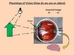
Light waves travel at a speed of 186000 miles (300 000kilometers) per second. Light is reflected into the eye by objects within the field of vision. White light is a combination of all the colours of the visual spectrum (rainbows). i.e., red, orange, yellow, green, blue, indigo, and violet. This can be demonstrated by passing white light through a glass prism which refracts or bends the rays of the different colors to a greater or lesser extent, depending on their wavelength. Red has the longest wavelength and violet the shortest.
SENSE OF SMELL
The nose has a dual function: respiration and sense of smell.
OLFACTORY NERVES (FIRST CRANIAL NERVES)
these are the sensory nerves of smell. They have their origin in special cells in the mucous membrane of the roof of the nose above the superior nasal conchae. On each side of the nasal septum nerve fibers from the cells pass through the cribriform plate of the ethmoid bone to the olfactory bulb, bundles of nerve fibers form the olfactory tract which passes backwards to the olfactory area in the temporal lobe of the cerebral cortex in each hemisphere where the impulses are interpreted and odour perceived.

PHYSIOLOGY OF SMELL
The sense of smell in human beings is generally less acute than in other animals. All odourous material give off chemical particles which are carried into the nose with the inhaled air and stimulate the nerve cells of the olfactory region when in solution in mucous. When an individual is continuously exposed to an odour, perception of the odour quickly decrease and eventually ceases. This loss of perception only affects that specific odour and adaptation probably occurs both in the cerebrum and in the nerve endings in the nose.
The air entering the nose is headed and convection currents carries eddies of inspired air from the main stream to the roof of the nose. Sniffing concentrates more particles more quickly in the roof of the nose. This increases the number of special cells stipulated and thus the perception of the smell. The sense of smell may affect the appetite. If the odour are pleasant the appetite may improve and vice versa.
Inflammation of the nasal mucosa prevents odorous substances from reaching the olfactory area of the nose, causing loss of the sense of smell. The usual is the common cold.

SENSE OF TASTE

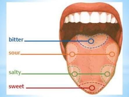
Taste buds are found in the papillae of the tongue and widely distributed in the epithelium of the tongue, soft palate, pharynx, and epiglottis. They consist of small bundles of cells and nerve endings of the glossopharyngeal, facial and vagus nerves (cranial nerves V11, 1X and X). Some of thr cells have hair-like microvilli on thier free border, projectibg towards tiny pores in the epithelium. The nerve cells are stimulated by chemical substance in solution that ente the pore. The nerve cell impulse are transmitted to thr thalamus then to the taste area in the cerebral cortex, one in each hemisphere, where taste is pwrceived. Four fundamental sensatuons of taste have been described; sweet, sour, bitter and salt. This is probably an oversimplification because perception varies widely and many 'tastes' cannot be easily classified. However, some tasres consistently stimulate taste buds in specific parts of thr tongue.
Sweet and salty, mainly at the tip
Sour, at the sides
Bitter, at the back
Hello @sirdeza
Thank you for this beautiful image you dedicated to space force health.

https://steemit.com/steemjet/@sirdeza/steemjetmedic
STEEMJET: On the wings of super star we are words and steem.
This has been educative
Downvoting a post can decrease pending rewards and make it less visible. Common reasons:
Submit
It's our work
Downvoting a post can decrease pending rewards and make it less visible. Common reasons:
Submit
I learnt a lot.
Downvoting a post can decrease pending rewards and make it less visible. Common reasons:
Submit
Loud it
Downvoting a post can decrease pending rewards and make it less visible. Common reasons:
Submit
Nice you could have added #steemit-education tag
Downvoting a post can decrease pending rewards and make it less visible. Common reasons:
Submit
Understanding man
I just did that
You have done noble
Downvoting a post can decrease pending rewards and make it less visible. Common reasons:
Submit
Cool....am learning also.
Downvoting a post can decrease pending rewards and make it less visible. Common reasons:
Submit
Superb and compelling...
Well done bro
Downvoting a post can decrease pending rewards and make it less visible. Common reasons:
Submit
Thank you boss
Downvoting a post can decrease pending rewards and make it less visible. Common reasons:
Submit
Steemjetmedic is here to stay, here to save and educate
Downvoting a post can decrease pending rewards and make it less visible. Common reasons:
Submit
Health is constant
Health is life
Downvoting a post can decrease pending rewards and make it less visible. Common reasons:
Submit
Concise comment!
Downvoting a post can decrease pending rewards and make it less visible. Common reasons:
Submit
Health is wealth.. This went a long way to refresh my memory.. Thank you
Downvoting a post can decrease pending rewards and make it less visible. Common reasons:
Submit
Aim archived
Downvoting a post can decrease pending rewards and make it less visible. Common reasons:
Submit
Welldone bro....u did justice to that topic....
Downvoting a post can decrease pending rewards and make it less visible. Common reasons:
Submit
As a physiologist you can't expect something less. Thank You for the complement
Downvoting a post can decrease pending rewards and make it less visible. Common reasons:
Submit
Bro this is a steemstem post.. Change one of the tags to steemstem and get a greater upvote
Downvoting a post can decrease pending rewards and make it less visible. Common reasons:
Submit
I just did that brother
Thank you for the light...
Downvoting a post can decrease pending rewards and make it less visible. Common reasons:
Submit
If you can make more of that, use steemstem tag and steemiteducation tag
Downvoting a post can decrease pending rewards and make it less visible. Common reasons:
Submit
This post has been rewarded with 30% upvote from @indiaunited-bot account. We are happy to have you as one of the valuable member of the community.
If you would like to delegate to @IndiaUnited you can do so by clicking on the following links: 5SP, 10SP, 15SP, 20SP 25SP, 50SP, 100SP, 250SP. Be sure to leave at least 50SP undelegated on your account.
to get community support and guidance
Please contribute to the community by upvoting this comment and posts made by @indiaunited.
Downvoting a post can decrease pending rewards and make it less visible. Common reasons:
Submit