Hello my steemit community,

In today's edition of IMPROVING HEALTH OF STEEMIANS, I want to talk to you about human ear, its anatomy and functions. This is somewhat of a prequel to my next posts. I will write about ear infections and I think its very important to know anatomy and functions of ear so you can fully understand diseases.
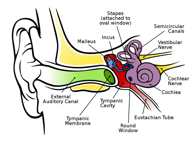
The ear is a multifaceted organ that connects the central nervous system to the external head and neck. This structure as a whole can be thought of as 3 separate organs:
OUTER EAR
MIDDLE EAR
INNER EAR
All three parts need to work in a collective to coordinate functions, such as hearing and balance.

source
OUTER EAR
The outer ear includes:
auricle- made up of cartilage confined by the skin. It is also known as the Ear pinna.
external auditory canal- formed by cartilage and bone. The canal measures about 4 cm in length.
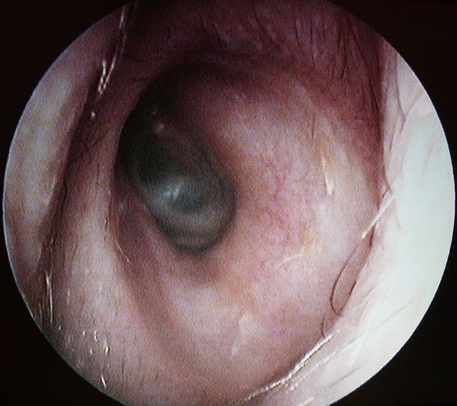
tympanic membrane- thin, concave membrane stretched across the inner end of the external auditory canal. It is also know as the eardrum. It marks the border between the outer ear and middle ear and serves as a transmitter of sound.
The external ear function is to act like an acoustic antenna that transmits sound waves to the sensitive middle ear structures. The auricle and the external auditory canal together form a unit that amplifies certain sound frequencies, typically in the range of 1000 to 4000 Hz. This amplification does not refer to an increase in the amplitude of the sound waves, but means that certain wave lengths vibrate better. Resonant frequency can be changed by different factors, such as cerumen and ear infections.
Two different acoustic paths are formed in the ear:
the direct path through the auditory canal
the indirect path through the helical shaped auricle
The asymmetric shape of the external auricle introduces delays in the path of sound for about 0.2 ms, which plays an important role in localizing the sound source. Therefore the loss of auricle does not cause significant acoustic damage, but it affects the ability of the person to localize the sound source in the space.
MIDDLE EAR
The middle ear is an air filled pit which communicates with the nasal cavity by a tube called Eustachian tube which allows the pressure to level between middle ear and throat. Eustachian tube is 3.5 cm long and two-thirds of are made of the cartilage, and the lateral one-third of the bone.
There are three bones in the middle ear:
malleus - attached to the eardrum
incus - the bridge bone between the malleus and the stapes
stapes- the smallest bone in the body
These three bones transmit the sound vibrations from the tympanic membrane into the inner ear which is the main function of the middle ear.
INNER EAR
The inner ear, also called the labyrinthine cavity, functions to conduct sound to the central nervous system, as well as to assist in balance.
The main components of the inner ear are:
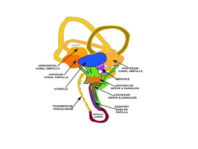
vestibule- the central structure in the inner ear. The outer wall of the vestibule contains the oval windows, which are the connection sites between the middle and inner ear. Internally, the vestibule contains two membranous sacs,
the utricle and the saccule (otolithic organs that sense linear acceleration in the horizontal and vertical planes). Within these organs are hair cells (the saccule macula and utricle macula) which are attached to nerve fibers. They serve as the vestibular (balance/equilibrium) sense organs.3 bony semicircular canals- each stands at right angles to the other 2 canals. They are filled with fluid and attached to cochlea and nerves. They send information on balance and head position to the brain.
(they sense rotational velocity in 3 dimensions)cochlea- spiral-shaped organ which is essential for hearing. It is split into 3 chambers :
scala vestibuli
scala media (contains organ of Corti)
scala tympani
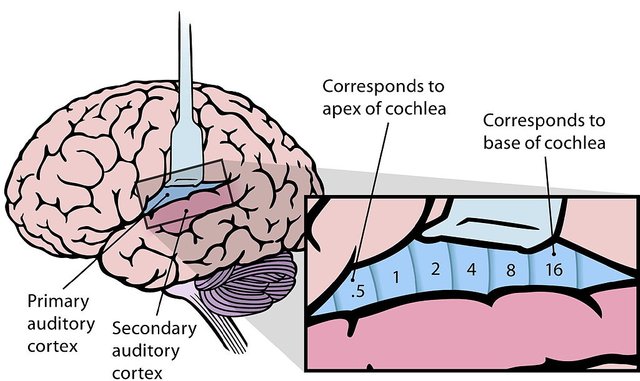
The organ of Corti contains hair cells and is the site of the conversion of sound waves into nerve impulses, which are sent to the brain.
PHYSIOLOGY OF HEARING
The auricle collects sound, and the external auditory tube conducts it to the eardrum. Under the influence of acoustic energy, the eardrum vibrates and transmits sound all the way to the stapes. Because of the difference in the surfaces of the stapes and the eardrum, the pressure ratio is 1:17. This means that the pressure on the stapes is 17 times greater than on the eardrum.
Because of that and some other anatomical differences, acoustic energy pressure increases 22 times at the entrance to the inner ear. Like the outer ear, and the middle ear has a resonant frequency, at about 1000 Hz.
In the inner ear (labyrinth) there is in-compressible fluid similar to liquor; perilymph in the scala tympani and the scala vestibuli, and endolymph in the scala media. Endolymph has a high positive potential (80–120 mV in the cochlea), relative to perilymph, due to its high concentration of positively charged ions.
The motion of the stapes causes the inner ear fluid to tremble. Sound waves are the converted into electrical impulses, which the auditory nerve sends to the brain. The brain then translates these electrical impulses as sound.

source
REFERENCE
Probst R., Basic Otorhinolaryngology, Thieme, 2006.
Marušić A., Anatomija čovjeka, Medicinska naklada, 2002.
Factual claims which are not sourced are from my own personal experience and med school.


I still remember learning the anatomy of inner ear. Not an easy task. :D
Downvoting a post can decrease pending rewards and make it less visible. Common reasons:
Submit
Definitely, one of the hardest part :) I remembered that prof. Jalšovec was asking me about inner ear, so I had to learn it so well :)
Downvoting a post can decrease pending rewards and make it less visible. Common reasons:
Submit
I can imagine, he demands pretty high level of knowledge from a first year student.
Downvoting a post can decrease pending rewards and make it less visible. Common reasons:
Submit
I am a doctor too... Very infomative article ...
Thanks for sharing @doctorcro
Good continuation
Downvoting a post can decrease pending rewards and make it less visible. Common reasons:
Submit
Thank you, its great to meet fellow doctor here :)
Downvoting a post can decrease pending rewards and make it less visible. Common reasons:
Submit
It's a shame we can only use certain types of pictures for steemstem posts, there are some really good images explaining the whole auditory process!
Also, some great videos on youtube xD doctor youtube, doctor google, and doctor Wikipedia are my best teachers so far in veterinarian school!!
But regerdless of that, great post doctorcro, you explained it very well!
Downvoting a post can decrease pending rewards and make it less visible. Common reasons:
Submit
Yes, its very hard to find enough good pictures. I never get the answer from authors of the pictures in time, so I find the free one :)
LOL, doctor wikipedia is everything
Downvoting a post can decrease pending rewards and make it less visible. Common reasons:
Submit
Next summer i'll probably learn some gimp or photoshop skills, they should make my posts better if i can work with them, getting an answer from the authors can take days or weeks... ain't nobody got time for that, still you found some really good pictures for this topic!! The most important part is the text imo, how well you write and how well you can "trap" your audience
Downvoting a post can decrease pending rewards and make it less visible. Common reasons:
Submit
Being A SteemStem Member
Downvoting a post can decrease pending rewards and make it less visible. Common reasons:
Submit