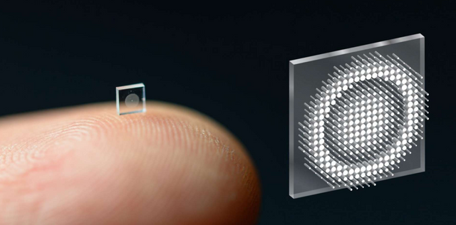
Recently, researchers have succeeded in creating a miniaturized, ultra-compact camera capable of taking high-definition and color pictures. By using a meta-surface rather than a conventional optic they have managed to reduce the camera to the size of a large grain of salt.
Miniaturized cameras could revolutionize medicine by making it much easier for doctors to see inside the human body. To that end, researchers at Princeton and Washington universities have succeeded in creating a camera the size of a grain of rice. Published in the journal Nature Communications, their paper details their metamaterial-based invention.
The researchers created a metasurface that captures light using 1.6 million optical nano-antennas. Each must have its unique geometry to properly form the optical wavefront. Designing the metasurface required complex simulations, using "massive amounts of memory and time," to test many different configurations of the nano-antennas.
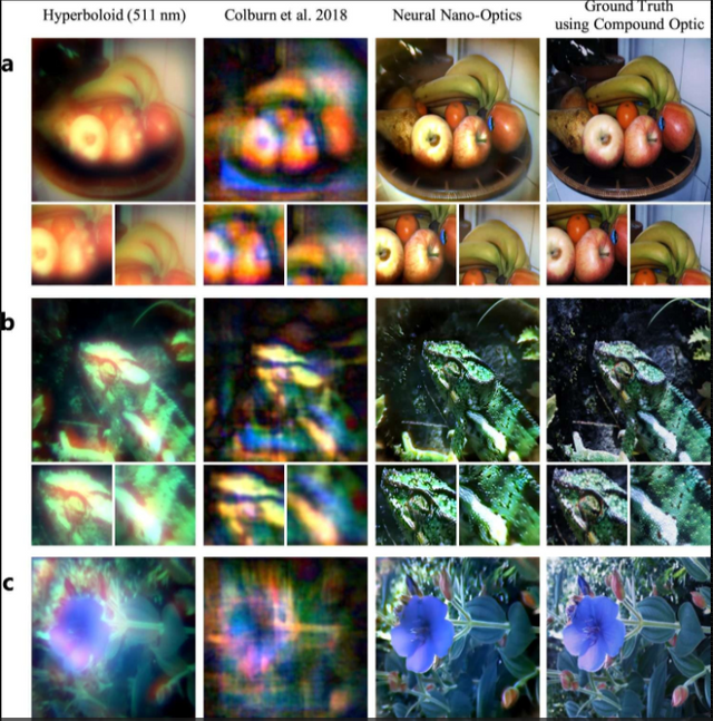
This image comparison is obtained using two other metamaterial-based cameras, the researchers' prototype, and a conventional camera. ©Princeton University.
A camera 550,000 times smaller than a conventional camera:
In addition to the simulations, the researchers created their own deconvolution algorithm to correctly reconstruct the resulting image. The result is a camera that measures only 0.5 mm thick and can take pictures with a similar quality to a conventional camera while being 550,000 times smaller in volume. This is not the first camera designed around a metasurface, but previous results were of much lower quality.
In addition to reducing the size of tools for endoscopy, the researchers envision using the same technique to turn surfaces into sensors. This would make it possible, for example, to convert the back shell of a smartphone into a giant camera and thus eliminate the traditional photo module.

